Erasmus MC Imaging Office medical image analysis
Application of in-house developed and open-source (automatic) image analysis methods for medical images (e.g. CT, MRI, etc.)
About
In the dynamic landscape of medical imaging, access to the latest imaging technologies and expertise is crucial. The Imaging Office of the Radiology & Nuclear Medicine department of the Erasmus MC offers a comprehensive range of services aimed at supporting all aspects related to medical imaging. From data storage to (software for) (automatic) image analysis to implementation in the clinic, the Imaging Office acts as a gateway to the combined knowledge and skills of the department. Even in an early stage of preparing a grant proposal in which imaging plays a role or for any imaging-related project, the Imaging Office is happy to advise on how to proceed.
We have access to a wide range of fantastic (AI) tools, developed by our own researchers. With the help of our experienced research software engineers, we can reuse these and other state-of-the-art open-source tools, to perform high quality image analysis.
Examples
In this section we provide you with some examples of digital image processing that can be performed, organized per body part. This list is not extensive and is growing every day, so don’t hesitate to contact us if your specific tool is not listed below!
Non-specific body part imaging
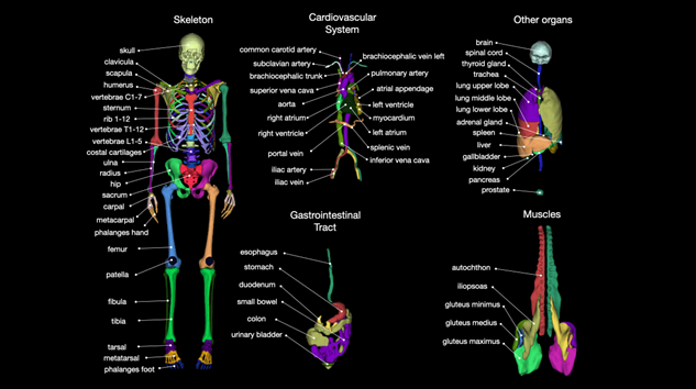
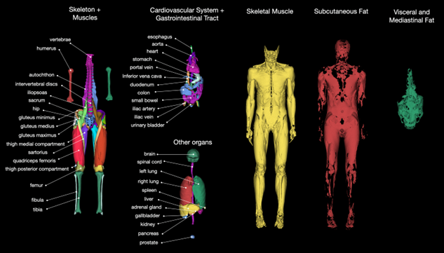
- Automatic organ segmentation on CT (see figure below)
- Automatic organ segmentation on MRI (see figure below)
- Automatic organ segmentation on a variety of modalities
- Minimally interactive soft-tissue tumor segmentation on MRI or CT
- Automatic tumor segmentation (and morphology measures, e.g. RECIST) on a variety of modalities
- Automatic radiomics analysis to develop prediction models on different modalities
- Automatic tumor quantification (e.g. diagnosis) using previously developed radiomics model (e.g. liver, soft tissue, rectal tumors) on MRI or CT
- Super-resolution method for multi-slice MRI
- Automatic registration for a variety of applications
Brain imaging
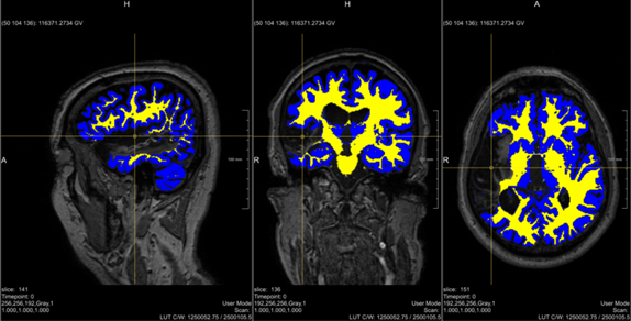
- Automatic brain (region) segmentation/volume on MRI
- Analysis of white matter tracts on brain diffusion MRI
- Arterial spin labelling (ASL) quantification on brain MRI
- White matter hyper intensities segmentation on brain MRI
- Automatic labelling of MRI scans to T1, T1C, T2, PD and derived maps
- Automatic glioma segmentation on MRI
- Multi-parametric analysis of the brain on (advanced) (PET-)MRI sequences
- Collateral scoring for stroke patients on CTA
- Brain vessel segmentation on CTA
- Automatic TICI scoring for stroke patients on DSA
- Brain atlas probability maps
Thoracic imaging
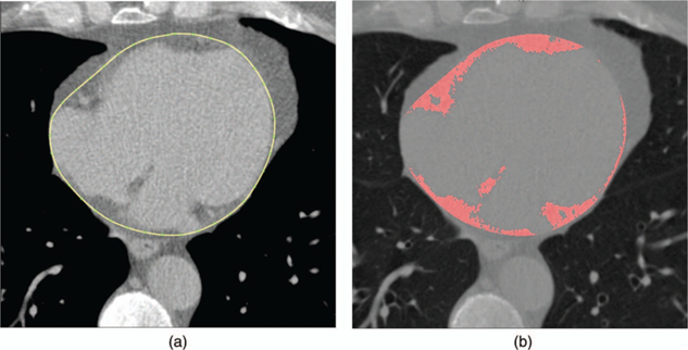
• Quantification of epicardial fat on CT
Musculoskeletal imaging
- Automatic bone age assessment on hand DXA scans
- Comprehensive knee (structures) segmentation on MRI
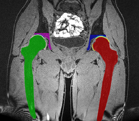
- Comprehensive hip (structures) segmentation on MRI
- Different biomarkers for a variety of hip disorders on MRI
(Collaborations with) other departments
The field of medical imaging is not restricted to the department of Radiology & Nuclear Medicine alone. Think of automatic 3D photocephalometric measurements of the skull for neurosurgery and skin cancer risk assessment on regular photography for dermatology. In ophthalmology there is for example automatic segmentation of cone photoreceptors in AO-FIO images, automatic endothelial cell counting, and a variety of manual and AI-assisted grading options for retinal imaging for quantification of disease burden (e.g. AMD) and morphology (e.g. vasculature, retinal layers) provided by the EyeNED Reading Center. In pulmonology, a variety of (semi-)automatic airway measurements and quantification of several structural lung diseases (e.g. CF, BPD) can be performed on lung CT scans by our colleagues from LungAnalysis. In the field of gynecology, automatic measurements can be performed on 3D ultrasound to assess fetus growth during pregnancy. From an epidemiology perspective, it is beneficial to use software to harmonize measurements for a very large number of study subjects, for example when measuring carotid and vertebrobasilar artery calcifications on CT scans. Finally, you could integrate for example pathology images in automatic diagnosis pipelines originally based on radiological images only.
Does one of these examples spark your interest? Don’t hesitate to contact the Imaging Office and we would be very happy to bring you in contact with the people involved!
Getting Started
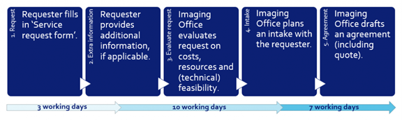
The Imaging Office works with a standardized service request form, on which can be indicated which services are requested with regard to imaging data, image analysis and image processing. The form can be submitted here. After reception of the fully completed request form, a confirmation of receipt will be sent within three working days and an intake interview will be scheduled within ten working days. On the basis of the intake interview it will be decided whether it is possible to accept the request.
Conditions of use
- The outcomes of any image analysis related request are very much dependent on the quality of the image data and the used algorithm. The Imaging Office cannot make any guarantees on final quality of the deliverables.
- If an image analysis pipeline is requested that has a low technology readiness level (TRE) the costs/timelines/outcomes are difficult to predict. The requester should be aware of this.
- If the project results in written deliverables (e.g. a master thesis or a publication), the work performed by the Imaging Office should be correctly referenced. If the work meets the criteria for co-authorship, the Imaging Office employee should be included as co-author. If it does not meet the criteria, the Imaging Office should be mentioned under acknowledgements. If the work consists of applying an imaging analysis pipeline, the publication on this pipeline should be referenced. If no publication exists, the pipeline developer should be mentioned under acknowledgements.
- A quality check on the delivered results is the responsibility of the requester (unless agreed upon differently) within three months.
Further information
Fair use and pricing
There will be costs associated with the service provided. These costs could be included in a grant application. An indication for the price can be requested via email.
Security controls
The following security controls are applicable:
- When data is being shared, a data transfer agreement (DTA) will be drafted
- When data is being processed, a data processing agreement (DPA) will be drafted
- Imaging data is only shared and processed after (pseudo-)anonymization